DICOM Anonymisation Software

DICOM Anonymisation Software

DICOM Anonymisation Software

DICOM anonymisation software is essential for the secure storage and sharing of medical images. It preserves patient privacy, ensures that personal data is anonymised and facilitates the use of images for teaching or research purposes without exposing sensitive patient information.

How do you choose the best DICOM anonymisation software? This article will discuss the key considerations for this.

What is DICOM anonymisation software?

DICOM anonymisation software protects medical images privacy by removing or replacing personal information within DICOM files.

DICOM anonymisation software’s goal is to make the data anonymous, allowing images to be used for medical or research purposes without risking patient privacy.

For example, when doctors share radiological images with colleagues, they must remove all identifiable information, such as the patient’s name, address, or other personal details.

How does it work?

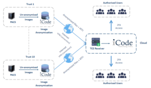
First, DICOM files are uploaded into the anonymisation software, which then scans the files for metadata tags containing personal information.

The software then removes or replaces this information with generic placeholders to protect the patient’s identity.

Pros of DICOM anonymisation software

DICOM anonymisation software is important to protect patients information especially with growing use of electronic health records that lead to more data being shared, increasing chances for accidental leaks of information.

Some benefits of DICOM anonymisation software include:

1. Compliance with regulations

Anonymising patient data is a legal requirement to protect patient confidentiality, so following strict privacy regulations, such as GDPR or HIPAA is essential.

2. Keeping patients’ information secure

Protecting medical information of the patients is more critical than ever, as cyberattacks have become sophisticated and can cause serious harm by stealing patients information or making it vulnerable to malicious actors.

3. Increase collaboration

Anonymised DICOM files help with collaboration across different departments, healthcare facilities, and research centers without compromising patient’s privacy.

4. Batch processing

Many DICOM anonymisation tools support batch processing, allowing the anonymisation of large sets of images at once (bulk DICOM anonymisation).

This feature is especially helpful for large healthcare facilities and research centers that handle substantial volumes of medical images.

Cons

Although bulk DICOM anonymisation software are very useful, the may have some cons:

1. Complex to use

Some DICOM anonymisation software can be complex to use, especially for healthcare professionals who are less technically proficient.

As a result, it may require some training, or having technical support to use DICOM anonymisation software properly without affecting the quality of patient care.

2. Potential for human error

Although anonymisation software can automate the process, there’s always a risk of human error.

For instance, a user may accidentally select the wrong settings, causing improper anonymisation, which results in unintentional patient data exposure.

3. Loss of data integrity

Sometimes anonymisation software can cause a loss of certain non-essential data that could be important in specific situations.

A clear example of this is when removing all patient-related data, it’s hard to trace an image back to the original patient for follow-up.

4. Not always 100% accurate

DICOM Anonymiser tools are very effective, although some advanced DICOM metadata might not be anonymised properly, which leaves some sensitive information exposed.

As a result, it may be essential to check the anonymisation process periodically to ensure patient privacy.

How to choose the right DICOM anonymisation software?

Choosing the best DICOM anonymisation software for your practice depends on many factors:

1. Ease of use

It’s preferable to look for a user-friendly software that can integrate easily into your workflow. As a healthcare provider, you should seek software with minimal learning time, ensuring ease of use without disrupting workflow.

2. Complaint with regulations

you have to ensure that the software you choose complies with privacy laws to avoid any legal problems and protect patient privacy.

3. Security features

DICOM anonymisation software varies in the security features, so you should make sure to choose the software that provides high protection of data during storage and transmission.

Some security features, such as encryption and secure cloud storage, provide great protection for patient privacy.

4. Scalability

The volume of the data may differ from healthcare facility to another, so you have to ensure that you have a software that can handle a large volume of data, whether now or later in case you need that.

This is very important especially for healthcare facilities or research centers that deal with a large amount of medical images and they sure need to protect their patient privacy.

5. Little errors

Although 100% accuracy is impossible, look for DICOM anonymisation software with minimal errors to ensure maximum security of patient information and prevent unintended data leaks.

6. Integration with other systems

A perfect DICOM anonymisation software should integrate well with other systems, such as the PACS system.

Best DICOM anonymisation software

PRIX Bulk DICOM Anonymiser is a powerful tool to bulk anonymise medical images and can integrate with PACS system.

It has some features that fit your need:

  • Effortless anonymisation: PIXR Bulk DICOM Anonymiser can anonymise large volumes of studies at once.
  • Multiple anonymisation profiles: It customizes with multiple profiles to meet the specific needs of different research, teaching, and clinical trials.
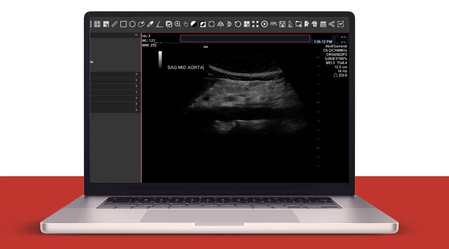
  • DICOM and pixel data anonymisation: It ensures precise anonymisation of the patient data in US images, maintaining data integrity and patient privacy with zero errors.
  • Integration with other systems: PRIX integrates with PACS, clinical trial servers, providing detailed log for each anonymisation step.

You can find what you need for anonymising your studies and cases perfectly in PIXR Bulk DICOM Anonymisation with its unique features.

FAQs

What is DICOM software?

DICOM software refers to specialized tools and applications designed to view, store, and share medical images in DICOM format, the standard format used in medical imaging.

How to anonymise DICOM files?

You can anonymise DICOM files using DICOM anonymisation software, as it preserves patient privacy while using images in teaching, research, or other uses.

PACS System Radiology

PACS System Radiology

PACS system radiology

PACS system radiology is a secure, reliable method to store, retrieve, and transport medical images. It provides benefits, such as remote access, and better-organized patient data.

They eliminate the need for hard-copy films and use digital images instead. This can help with providing better patient care and making more accurate diagnoses, especially in radiology.

What is PACS?

PACS stands for Picture Archiving and Communication System. It securely transports medical imaging information, allowing healthcare professionals to store and transmit images and clinical reports.

How does it work?

The PACS system stores and transfers medical images in DICOM (Digital Imaging and Communications in Medicine) format and supports several functions:

  • Image acquisition: Medical images are digitally captured from multiple imaging methods, such as X-ray, CT, and MRI, adhering to DICOM standards.
  • Image transfer and storage: These digital images are sent to the PACS server that stores and archives them in a central database.
  • Image distribution and viewing: Radiologists can access and view stored images from any PACS workstation connected to the network.
  • Reporting: Radiologists can create and save diagnostic reports with their images in the PACS.
  • Integration: PACS systems can integrate with other systems, such as RIS (Radiology Information System).

PACS and RIS

PACS can integrate with the Radiology Information System (RIS), enabling bi-directional communication between the systems. RIS sends patient data to PACS, and PACS returns acquired images and reports, allowing a coordinated radiology workflow.

Importance of PACS in radiology

Radiologists benefit from PACS in many ways, including:

  • Remote access: PACS enables radiologists to access diagnostic images anytime, improving diagnosis accuracy, speed, and healthcare quality.
  • Film archives replacement: PACS eliminates the need for film archives, reduces physical storage requirements, and promotes efficient cooperation across departments.
  • Easy for radiologists: Radiologists can use the PACS system to manage their work. They take the necessary images, review them in the workstation, sending them to the digital archive, and the images are now available for healthcare professionals who have access to the PACS system.

Types of PACS systems

Types of PACS systems include:

1. Traditional PACS

Traditional PACS represents the cornerstone of image storage, retrieval, and distribution within a local network.

They offer strong control and privacy but it’s limited by physical space and requires continuous maintenance.

Traditional PACS are perfect for facilities that prefer keeping all operations in-house and have the necessary infrastructure.

2. Cloud-based PACS

They are more modern than traditional PACS, as they benefit from the internet for image storage and management. The images can be accessed from any device, at any time without great concern about physical storage limitations or maintenance.

They offer scalable storage solutions, usually requiring a subscription.

3. Hybrid systems

Hybrid systems blend traditional and cloud-based PACS. They offer the best of both systems:

  • Control and Security
  • flexibility and scalability of the cloud

They are fit for those looking forward to maintaining on-site backup while taking advantage of cloud storage as well.

Benefits of PACS system radiology

Medical PACS systems can provide a lot of benefits, especially in the radiology field. Some of these benefits include:

  • More efficient diagnoses
  • Reduced costs
  • High-quality medical images
  • Instant remote access for medical images
  • Increased integration between departments and facilities
  • Improve patient care
  • Ease of use and scalability of cloud-based PACS

For more details about some of the benefits:

  • Reduced costs: Digital storage is generally less expensive than storage needed for hard-copy films, which makes the PACS system a cost-effective solution.
  • Better patient data management: All patient information can be accessed through a single point of access as images are integrated with the radiology information system.
  • Faster workflow: There’s no need for manual transport and retrieval of images. It’s a rapid process with the help of the PACS system, as radiologists can access images quickly and provide better patient care.

Best PACS system radiology

The best PACS system according to the number of installs includes:

  1. GE Healthcare – Centricity
  2. Change Healthcare – Radiology Solutions
  3. FUJIFILM Medical Systems – Synapse
  4. IBM – Merge PACS
  5. Philips Healthcare – iSite
  6. Agfa Healthcare – Impax
  7. Novarad – NovaPACS
  8. Sectra – SectraPACS
  9. Carestream Health – Carestream vue
  10. Change Healthcare

PACS system radiology cost factors

How much does a PACS system cost? There are many factors affecting the cost of the PACS system, such as:

  • Total storage capacity: Solid-state storage (SSD) is more available and much faster to access than spinning disks (HDD). SSD costs about double the similar capacity of disk storage.
  • Maintenance and support: The PACS system requires annual maintenance and support which may affect the cost of the PACS system.
  • Type of PACS system used: Also choosing the appropriate type of PACS system for you may influence the total cost.

FAQs

What is the PACS system in radiology?

PACS system is a secure method to store and transport medical images according to DICOM format. It helps radiologists to access diagnostic images anywhere, sharing images with other healthcare professionals, and providing better patient care.

What are the main components of PACS?

PACS system for radiology components include:

  • Main server: The main server is the most important part of a PACS. It includes the database structure, RIS interface, web servers, and other image distribution servers and interfaces.
  • Database: This refers to the place at which all the important information is stored including all the data related to the patient study and examination.
  • Health level seven (HL7): The HL7 is the part that receives all the info caused by RIS and then forwards it to the PACS.
  • Imaging modalities: These are machines used to capture digital medical images, such as X-ray machines, MRI machines, and Ultrasound equipment.
  • Workstations: These are computers equipped with DICOM viewers that allow radiologists to access, view, and interpret stored images.
  • Storing archives: DICOM images are stored in the servers or cloud storage, either on-site, in the cloud, or both (hybrid).

A secure network connection is also necessary for the healthcare PACS system to work efficiently.

Rosenfield Exhibiting at UKIO 2024

News

Rosenfield Exhibiting at UKIO 2024

Rosenfield at UKIO 2024

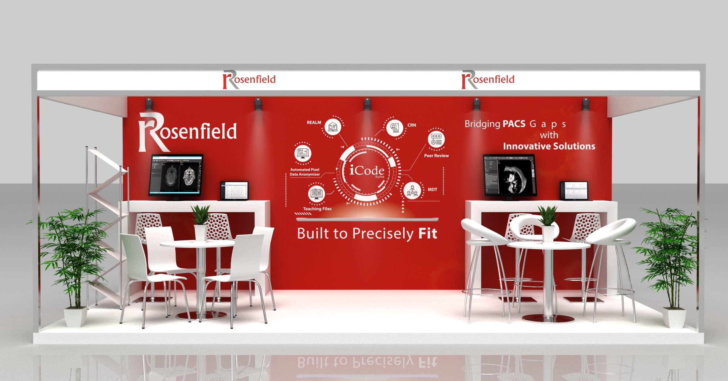
We’re thrilled to announce that we will be exhibiting at hashtagUKIO24!

Join us to get hands-on with our latest innovative solutions and dive into our interactive demos! Our team is ready to meet you and discuss how our products can empower your workflow. 

Mark Your Calendars:
Date: 10,11,12 June, 2024
Location: ACC, Liverpool
Booth: A5/10


Fill out this form to schedule your Demo during the event:
https://lnkd.in/drKPmvwu

We can’t wait to see you there and explore the possibilities together

#UKIO #Medical_imaging #HealthcareIT #Oncology #Radiology  

3 Tips to Organise an Effective Tumour Board

3 Tips to Organise An Effective Tumour Board

Blog

3 Tips to Organise An Effective Tumour Board

Enhancing Cancer Care: 3 Tips to Organise an Effective Tumour Board

Introduction: As we mark World Cancer Month, it’s a timely reminder of the global impact of Cancer and the importance of effective collaboration in combating this disease. It’s important to reflect on the critical role that well-organised Tumour Board meetings play in enhancing cancer care efficiency. These multidisciplinary gatherings bring together healthcare professionals from various specialties to collectively review complex cases, discuss treatment options, and formulate comprehensive care plans for cancer patients. By fostering collaboration, innovation, and evidence-based decision-making, these Board meetings contribute significantly to improving patient outcomes and quality of life. In this article, we’ll explore three tips to organise Tumour Boards effectively and enhance cancer care outcomes. 

1. Establish Clear Objectives and Agendas

Set clear objectives and agendas for each meeting to ensure focus and productivity. Define specific goals, such as reviewing diagnostic imaging, discussing treatment options, and developing individualised care plans.

2. Enhance Data Accessibility and Integration

Access to comprehensive patient data is crucial in discussions and decision-making. Integrate electronic health records (EHRs), imaging studies, pathology reports, and other relevant information into a unified platform. By centralizing data access, healthcare professionals can make more informed decisions, reduce duplication of efforts, and improve patient outcomes.

3. Ensure All Images Are Reviewed Prior to the Tumour Board

Comprehensive imaging studies are a cornerstone of cancer diagnosis and treatment planning. It is also one of the standards the RCR addressed in their recent article. To maximize the effectiveness of Tumour Board meetings, it’s essential to ensure that all relevant images are thoroughly reviewed beforehand. This includes radiographs, CT scans, MRIs, PET scans, and any other imaging modalities pertinent to the patient’s case. By meticulously examining these images prior to the meeting, healthcare professionals can identify key findings, gain a deeper understanding of the patient’s condition and save time during the meeting.

In today’s digital age, leveraging technology can significantly enhance the efficiency and effectiveness of Tumour Board meetings. With solutions like iCode MDT, healthcare professionals can streamline the entire process, from adding comments prior to the meeting to documenting patient data and treatment plans. By using iCode MDT’s intuitive platform, organizers can create customized agendas, assign tasks, and track progress seamlessly. Additionally, iCode MDT facilitates real-time collaboration and communication among team members, ensuring that everyone is on the same page and working towards common goals. Incorporating technology like iCode MDT into Tumour Board meetings not only saves time and resources but also improves overall outcomes for cancer patients.  

Conclusion: Organising Tumour Boards effectively is essential in enhancing cancer care and improving patient outcomes. By fostering reviewing images, establishing clear objectives and enhancing data accessibility, healthcare professionals can maximize the impact of MDT meetings. Leveraging innovative solutions like iCode MDT further enhances collaboration, streamlines processes, and ultimately contributes to better cancer care delivery. Together, let’s continue to work towards a future where cancer is no longer a life-threatening disease, but a manageable condition with improved treatment options and outcomes. 

Rosenfield Health’s Innovative Partnership with East of England Academy

News

Rosenfield Health Tech and East of England Academy Forge Pioneering Partnership in Advancing Imaging Education

Jan 11, 2024 | Radiology Education | United Kingdom

Cardiff, UK – January 2024 
In a ground-breaking collaboration poised to redefine imaging education, Rosenfield Health Tech, a leading provider of radiology education solutions is thrilled to announce its partnership with the East of Radiology England Academy. This collaboration marks the biggest deployment of our revolutionary iCode Teaching Files Solution, designed to elevate medical imaging education standards, in PACS across the UK. 

Revolutionizing Medical Education with iCode Teaching Files Solution 

Rosenfield Health's Innovative Partnership with East of England Academy

Rosenfield Health Tech‘s iCode Teaching Files Solution represents a paradigm shift in optimizing teaching studies and materials. Adhering to ACR standard, our product is specifically crafted to enhance the learning experience by anonymizing and archiving compelling studies on a dedicated server. The separate database can be accessed locally or regionally, providing unparalleled accessibility for radiologists.

Our vendor-neutral solution will be seamlessly integrated with PACS from five different vendors. This adaptability ensures a smooth integration process for medical institutions, regardless of their existing systems, contributing to a cohesive and streamlined educational experience. 

Unprecedented Reach: 31 NHS Trusts in the UK.

Rosenfield Health's Innovative Partnership with East of England Academy

This collaboration will expand Rosenfield Health’s installed base to 31 NHS trusts in the UK, up from 18. By deploying our iCode TFS in these trusts, we aim to enhance the quality and efficiency of imaging education, and to foster a culture of continuous learning and improvement among medical imaging professionals.

Hameed Rafiee, Virtual Imaging Academy Development Lead, NHS said: “As Virtual Imaging Academy Development Lead for the East of England, I am delighted to announce our partnership with Rosenfield Health to deploy their fantastic iCode teaching solution across the East of England for the purpose of developing imaging education material for radiologists, radiographers, sonographers, junior doctors, medical students, and any other professional group with an imaging learning need. This partnership will enable us to revolutionise and accelerate imaging education across the region, thereby improving training quality, reducing the burden on trainers and aiding workforce retention. 

Shaping the Future of Medical Education in the UK 

The contract was signed in December 2023 for an initial 2-year term and outlines a strategic roadmap to be completed within six months.

Grahame Penny, Senior Commercial Lead (UK/I) GE HealthCare, expresses his enthusiasm for leading the delivery of this transformative project: “We are very excited to lead the delivery of this project. We have a strong relationship with Rosenfield and have a great track record of delivering projects together successfully and on time. This partnership with the Academies for East of England will bring major benefits for clinicians (and future patient care) and will continue to strengthen our collaboration in the future.” 

Ahmed Adnan Elsharkawy, Managing Director, Rosenfield Health Tech said:We are excited to be awarded this partnership with the East of England Academy, a prestigious institution that shares our vision of advancing medical imaging education through cutting-edge technology. We believe that this project will not only benefit the medical imaging community in the UK, but also inspire other regions and countries to adopt similar solutions for their own education needs. 

This partnership underscores Rosenfield Health Tech‘s dedication to providing healthcare professionals with the tools they need to excel, contributing to enhanced patient care. Stay tuned as we set new standards for imaging education in the ever-evolving radiology sector.