Radiology Teaching Files

Radiology Teaching Files - Rosenfiled Health

Radiology Teaching Files

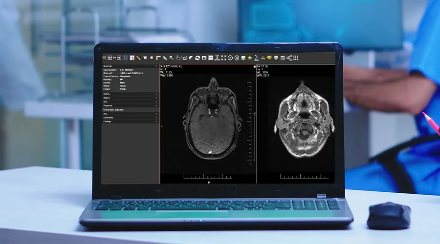
Radiology teaching files are powerful tools for radiologists. They allow the archiving of interesting cases to ensure important findings are not missed while also enhancing diagnostic skills.

These teaching files serve multiple purposes, whether it’s for educating radiology students, discussing cases with colleagues, contributing to the medical community, or keeping up with effective e-learning methods for radiology.

What Are Teaching Files?

Radiology teaching files are collections of clinical cases with teaching value.

These files provide real examples of radiology cases that may help with teaching purposes, whether for radiologists or radiology students.

Generally, radiology education relies on studying and referencing previous patient cases but storage, accessibility, and shareability with students can present challenges. Additionally, studies should be anonymized.

So, radiology teaching files can serve several purposes, such as:

  • To ensure important findings are not missed.
  • Archiving cases for clinical follow-up.
  • As a reference for better understanding diseases.
  • Facilitating secure Knowledge transfer and assessment.
  • Enabling quick retrieval of anonymized patient cases.

Structure Of Radiology Teaching Files

Radiology teaching files are organized to make them easy for learners to navigate and understand. Each case typically includes:

  • Patient information: This Includes the patient’s age, gender, medical history, and symptoms.
  • Imaging studies: X-rays, CT scans, MRIs, or ultrasounds are often presented in multiple views to better understand the cases.
  • Annotations and labels: Key areas of the image are often highlighted to direct the learners to a significant finding.
  • Diagnosis and discussion: A detailed explanation of the diagnosis, including why certain findings on the images led to that conclusion.
  • Teaching points: Summary points that highlight important lessons from the case.

Teaching Files Types

Teaching files can be divided into three types:

1. Personal Teaching Files

Personal teaching files are for individual use. The owner may have an interest in a very specific case for quality control, reviewing his work, teaching, or clinical follow-up.

2. Shared In-House Teaching Files

Personal teaching files can be migrated into a shared teaching file environment. Additional information can be added to make the case available for colleagues to view and learn from.

3. Public Teaching Files

They are built on a shared teaching files model but contain more comprehensive content. They may undergo a formal review before publication and sometimes require a subscription fee.

Teaching File Case Types

Teaching file case types include:

1. Cardiac

Cardiac teaching files focus on heart-related cases and cardiovascular conditions. They include imaging for:

  • coronary artery disease
  • congenital heart anomalies
  • heart failure
  • valvular diseases
  • others

Radiologists can use these files for better understanding of heart structure and function. Files can be also helpful for assessment of cardiac conditions.
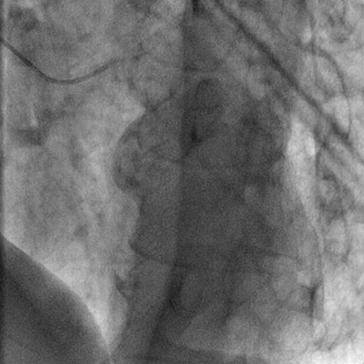
Cardiac and Interventional teaching files

2. Chest

Chest teaching files cover diseases of the chest, such as:

  • pneumonia.
  • chest tumors.
  • interstitial lung diseases.
  • pleural abnormalities.

They help radiologists enhance their skills in chest cases.

3. Gastrointestinal

Gastrointestinal teaching files focus on the digestive system including the esophagus, stomach, intestines, liver, pancreas, and biliary system.

They include cases of various gastrointestinal diseases, such as:

  • Liver diseases
  • Colorectal cancers
  • pancreatitis
  • bowel obstruction

Radiologists can archive these teaching files for educational purposes or to enhance their skills.

4. Genitourinary

These files focus on the urinary and reproductive systems. Files can include:

  • Renal cell carcinoma
  • Polycystic kidney disease
  • Urinary tract infections
  • Urinary stones
  • Hydronephrosis
  • Ovarian masses

These files help radiologists assess urological structure and functions and improve their experience in the diagnosis of urinary tract masses and pathologies.
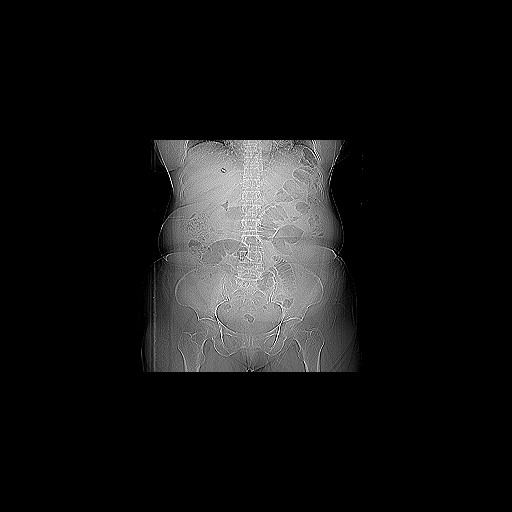
CT- Genitourinary teaching files

5. Interventional

Interventional teaching files include some procedures, such as:

  • Biopsies
  • Catheter placements
  • Angioplasty
  • Tumor ablation

These cases help radiologists learn more about the procedures and improve patient management skills.

6. Mammography

Mammography files specialize in breast imaging cases, such as:

  • Breast tumors
  • Fibrocystic changes
  • Breast cysts
  • Ductal carcinoma in situ

Radiologists can gain valuable experience in breast cases through mammography teaching files.
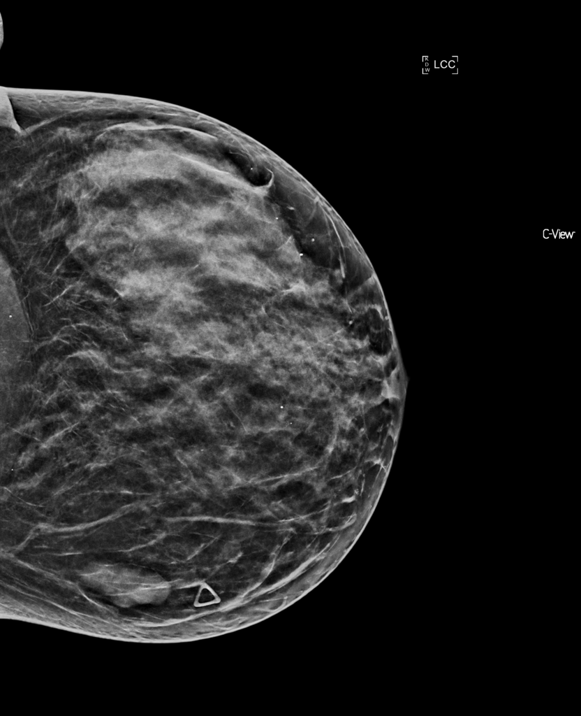
Mammography teaching files

7. Musculoskeletal

Musculoskeletal teaching files focus on improving radiologist’s skills in recognizing bone and soft tissue abnormalities, such as:

  • Osteoarthritis
  • Soft tissue tumors
  • Fractures
  • Bone tumors
  • Sports-related injuries

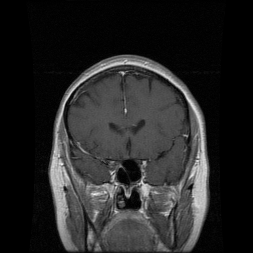
8. Neuroradiology

Neuroradiology files guide radiologists in recognizing brain abnormalities and how to differentiate normal from abnormal brain or nervous structure. They cover cases, such as:

  • Stroke
  • Brain tumors
  • Multiple Sclerosis
  • Traumatic brain injuries

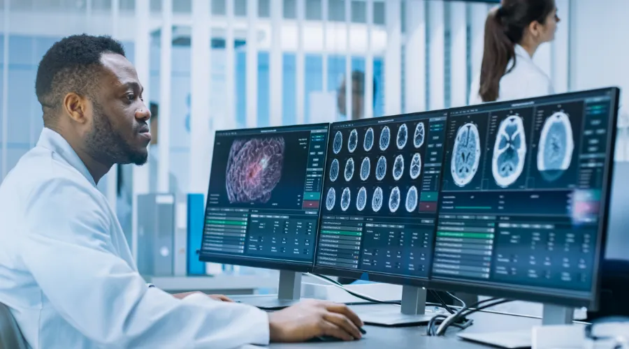
MRI Neuroradiology teaching files

9. Nuclear Medicine

These files cover radiotracer use, interpretation of uptake patterns, and correlation with clinical scenarios. They provide cases like:

  • Thyroid nodules
  • Bone metastases
  • Neuroendocrine tumors

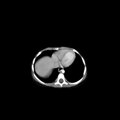
10. Pediatrics

Pediatric files train radiologists to recognize specific conditions in children. Pediatric cases often include:

  • Rickets
  • Congenital malformations
  • Neuroblastoma
  • Pediatric trauma
  • Respiratory conditions

Pediatric teaching files

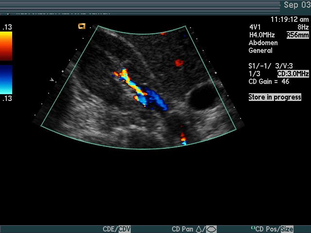
11. Ultrasound

Ultrasound case studies are essential for helping radiologists identify numerous conditions and enhance their diagnostic skills. They may include cases on:

  • Gallstones
  • Liver disease
  • Obstetric and gynecologic conditions
  • Thyroid nodules
  • Abdominal conditions
  • Vascular conditions

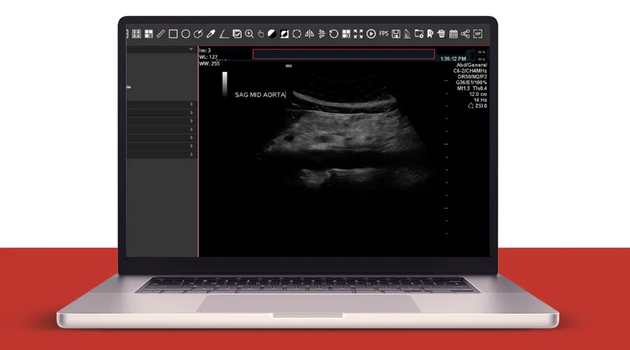
Ultrasound teaching files

Importance Of Teaching Files For Radiologists

Radiologists may be advised to build their teaching files for the following reasons:

1. Increase Your Expertise

Establishing teaching files encourages radiologists to engage with cases on a deeper level.

It fosters critical thinking in analyzing images, making accurate diagnoses, and explaining findings.

This process sharpens diagnostic skills and enhances a radiologist’s level of experience.

2. Education Purposes

Radiology teaching files enrich the medical community by providing real and often interesting cases. This helps medical students, radiologists, and colleagues. Importantly, difficult cases recorded in teaching files will be more easily diagnosed in the future.

3. Self-Learning And Review

Teaching files are not only for the broader medical community but can also serve as a valuable personal learning resource. Having cases documented allows radiologists to review challenging cases, rare pathologies, or those with subtle findings.

iCode Teaching Files

iCode Teaching Files is a web teaching files solution that helps radiologists archive their interesting studies and build imaging libraries according to ACR standards.

It archives interesting cases on a separate server with a separate database. You can keep the radiology teaching archive safe when the PACS vendor is changed.

Examples of iCode teaching files features include:

  • Easy file search and retrieval
  • Robust data anonymisation
  • Ability to export studies with several media files, such as presentations, videos, JPEG, TIFF & DICOM..etc
  • Manage teaching events easily with a streamlined workflow and lectures bank to enhance learning
  • Building scalable teaching archive across specific region or country.

iCode Teaching Files Radiology Exam System

The iCode Teaching File Solution is revolutionizing radiology education with its comprehensive exam module, designed to enhance both teaching and learning.

This system assists radiologists in creating radiology exams for their students. The exam module in iCode Teaching Files allows radiologists to craft multiple-choice questions (MCQs) based on archived medical images.

It also enables educators to share quiz links with their students, allowing them to take exams directly through the teaching files system.

With detailed analytics and instant feedback, both learners and educators can track progress and identify areas for improvement, ultimately elevating the quality of training.

iCode offers a rich set of features tailored to meet your educational needs, enabling you to archive cases quickly and effortlessly. Additionally, its comprehensive exam module supports and enhances the educational process.


DICOM Anonymisation Software

DICOM Anonymisation Software

DICOM Anonymisation Software

DICOM anonymisation software is essential for the secure storage and sharing of medical images. It preserves patient privacy, ensures that personal data is anonymised and facilitates the use of images for teaching or research purposes without exposing sensitive patient information.

How do you choose the best DICOM anonymisation software? This article will discuss the key considerations for this.

What is DICOM anonymisation software?

DICOM anonymisation software protects medical images privacy by removing or replacing personal information within DICOM files.

DICOM anonymisation software’s goal is to make the data anonymous, allowing images to be used for medical or research purposes without risking patient privacy.

For example, when doctors share radiological images with colleagues, they must remove all identifiable information, such as the patient’s name, address, or other personal details.

How does it work?

First, DICOM files are uploaded into the anonymisation software, which then scans the files for metadata tags containing personal information.

The software then removes or replaces this information with generic placeholders to protect the patient’s identity.

Pros of DICOM anonymisation software

DICOM anonymisation software is important to protect patients information especially with growing use of electronic health records that lead to more data being shared, increasing chances for accidental leaks of information.

Some benefits of DICOM anonymisation software include:

1. Compliance with regulations

Anonymising patient data is a legal requirement to protect patient confidentiality, so following strict privacy regulations, such as GDPR or HIPAA is essential.

2. Keeping patients’ information secure

Protecting medical information of the patients is more critical than ever, as cyberattacks have become sophisticated and can cause serious harm by stealing patients information or making it vulnerable to malicious actors.

3. Increase collaboration

Anonymised DICOM files help with collaboration across different departments, healthcare facilities, and research centers without compromising patient’s privacy.

4. Batch processing

Many DICOM anonymisation tools support batch processing, allowing the anonymisation of large sets of images at once (bulk DICOM anonymisation).

This feature is especially helpful for large healthcare facilities and research centers that handle substantial volumes of medical images.

Cons

Although bulk DICOM anonymisation software are very useful, the may have some cons:

1. Complex to use

Some DICOM anonymisation software can be complex to use, especially for healthcare professionals who are less technically proficient.

As a result, it may require some training, or having technical support to use DICOM anonymisation software properly without affecting the quality of patient care.

2. Potential for human error

Although anonymisation software can automate the process, there’s always a risk of human error.

For instance, a user may accidentally select the wrong settings, causing improper anonymisation, which results in unintentional patient data exposure.

3. Loss of data integrity

Sometimes anonymisation software can cause a loss of certain non-essential data that could be important in specific situations.

A clear example of this is when removing all patient-related data, it’s hard to trace an image back to the original patient for follow-up.

4. Not always 100% accurate

DICOM Anonymiser tools are very effective, although some advanced DICOM metadata might not be anonymised properly, which leaves some sensitive information exposed.

As a result, it may be essential to check the anonymisation process periodically to ensure patient privacy.

How to choose the right DICOM anonymisation software?

Choosing the best DICOM anonymisation software for your practice depends on many factors:

1. Ease of use

It’s preferable to look for a user-friendly software that can integrate easily into your workflow. As a healthcare provider, you should seek software with minimal learning time, ensuring ease of use without disrupting workflow.

2. Complaint with regulations

you have to ensure that the software you choose complies with privacy laws to avoid any legal problems and protect patient privacy.

3. Security features

DICOM anonymisation software varies in the security features, so you should make sure to choose the software that provides high protection of data during storage and transmission.

Some security features, such as encryption and secure cloud storage, provide great protection for patient privacy.

4. Scalability

The volume of the data may differ from healthcare facility to another, so you have to ensure that you have a software that can handle a large volume of data, whether now or later in case you need that.

This is very important especially for healthcare facilities or research centers that deal with a large amount of medical images and they sure need to protect their patient privacy.

5. Little errors

Although 100% accuracy is impossible, look for DICOM anonymisation software with minimal errors to ensure maximum security of patient information and prevent unintended data leaks.

6. Integration with other systems

A perfect DICOM anonymisation software should integrate well with other systems, such as the PACS system.

Best DICOM anonymisation software

PRIX Bulk DICOM Anonymiser is a powerful tool to bulk anonymise medical images and can integrate with PACS system.

It has some features that fit your need:

  • Effortless anonymisation: PIXR Bulk DICOM Anonymiser can anonymise large volumes of studies at once.
  • Multiple anonymisation profiles: It customizes with multiple profiles to meet the specific needs of different research, teaching, and clinical trials.
  • DICOM and pixel data anonymisation: It ensures precise anonymisation of the patient data in US images, maintaining data integrity and patient privacy with zero errors.
  • Integration with other systems: PRIX integrates with PACS, clinical trial servers, providing detailed log for each anonymisation step.

You can find what you need for anonymising your studies and cases perfectly in PIXR Bulk DICOM Anonymisation with its unique features.

FAQs

What is DICOM software?

DICOM software refers to specialized tools and applications designed to view, store, and share medical images in DICOM format, the standard format used in medical imaging.

How to anonymise DICOM files?

You can anonymise DICOM files using DICOM anonymisation software, as it preserves patient privacy while using images in teaching, research, or other uses.

PACS System Radiology

PACS System Radiology

PACS system radiology

PACS system radiology is a secure, reliable method to store, retrieve, and transport medical images. It provides benefits, such as remote access, and better-organized patient data.

They eliminate the need for hard-copy films and use digital images instead. This can help with providing better patient care and making more accurate diagnoses, especially in radiology.

What is PACS?

PACS stands for Picture Archiving and Communication System. It securely transports medical imaging information, allowing healthcare professionals to store and transmit images and clinical reports.

How does it work?

The PACS system stores and transfers medical images in DICOM (Digital Imaging and Communications in Medicine) format and supports several functions:

  • Image acquisition: Medical images are digitally captured from multiple imaging methods, such as X-ray, CT, and MRI, adhering to DICOM standards.
  • Image transfer and storage: These digital images are sent to the PACS server that stores and archives them in a central database.
  • Image distribution and viewing: Radiologists can access and view stored images from any PACS workstation connected to the network.
  • Reporting: Radiologists can create and save diagnostic reports with their images in the PACS.
  • Integration: PACS systems can integrate with other systems, such as RIS (Radiology Information System).

PACS and RIS

PACS can integrate with the Radiology Information System (RIS), enabling bi-directional communication between the systems. RIS sends patient data to PACS, and PACS returns acquired images and reports, allowing a coordinated radiology workflow.

Importance of PACS in radiology

Radiologists benefit from PACS in many ways, including:

  • Remote access: PACS enables radiologists to access diagnostic images anytime, improving diagnosis accuracy, speed, and healthcare quality.
  • Film archives replacement: PACS eliminates the need for film archives, reduces physical storage requirements, and promotes efficient cooperation across departments.
  • Easy for radiologists: Radiologists can use the PACS system to manage their work. They take the necessary images, review them in the workstation, sending them to the digital archive, and the images are now available for healthcare professionals who have access to the PACS system.

Types of PACS systems

Types of PACS systems include:

1. Traditional PACS

Traditional PACS represents the cornerstone of image storage, retrieval, and distribution within a local network.

They offer strong control and privacy but it’s limited by physical space and requires continuous maintenance.

Traditional PACS are perfect for facilities that prefer keeping all operations in-house and have the necessary infrastructure.

2. Cloud-based PACS

They are more modern than traditional PACS, as they benefit from the internet for image storage and management. The images can be accessed from any device, at any time without great concern about physical storage limitations or maintenance.

They offer scalable storage solutions, usually requiring a subscription.

3. Hybrid systems

Hybrid systems blend traditional and cloud-based PACS. They offer the best of both systems:

  • Control and Security
  • flexibility and scalability of the cloud

They are fit for those looking forward to maintaining on-site backup while taking advantage of cloud storage as well.

Benefits of PACS system radiology

Medical PACS systems can provide a lot of benefits, especially in the radiology field. Some of these benefits include:

  • More efficient diagnoses
  • Reduced costs
  • High-quality medical images
  • Instant remote access for medical images
  • Increased integration between departments and facilities
  • Improve patient care
  • Ease of use and scalability of cloud-based PACS

For more details about some of the benefits:

  • Reduced costs: Digital storage is generally less expensive than storage needed for hard-copy films, which makes the PACS system a cost-effective solution.
  • Better patient data management: All patient information can be accessed through a single point of access as images are integrated with the radiology information system.
  • Faster workflow: There’s no need for manual transport and retrieval of images. It’s a rapid process with the help of the PACS system, as radiologists can access images quickly and provide better patient care.

Best PACS system radiology

The best PACS system according to the number of installs includes:

  1. GE Healthcare – Centricity
  2. Change Healthcare – Radiology Solutions
  3. FUJIFILM Medical Systems – Synapse
  4. IBM – Merge PACS
  5. Philips Healthcare – iSite
  6. Agfa Healthcare – Impax
  7. Novarad – NovaPACS
  8. Sectra – SectraPACS
  9. Carestream Health – Carestream vue
  10. Change Healthcare

PACS system radiology cost factors

How much does a PACS system cost? There are many factors affecting the cost of the PACS system, such as:

  • Total storage capacity: Solid-state storage (SSD) is more available and much faster to access than spinning disks (HDD). SSD costs about double the similar capacity of disk storage.
  • Maintenance and support: The PACS system requires annual maintenance and support which may affect the cost of the PACS system.
  • Type of PACS system used: Also choosing the appropriate type of PACS system for you may influence the total cost.

FAQs

What is the PACS system in radiology?

PACS system is a secure method to store and transport medical images according to DICOM format. It helps radiologists to access diagnostic images anywhere, sharing images with other healthcare professionals, and providing better patient care.

What are the main components of PACS?

PACS system for radiology components include:

  • Main server: The main server is the most important part of a PACS. It includes the database structure, RIS interface, web servers, and other image distribution servers and interfaces.
  • Database: This refers to the place at which all the important information is stored including all the data related to the patient study and examination.
  • Health level seven (HL7): The HL7 is the part that receives all the info caused by RIS and then forwards it to the PACS.
  • Imaging modalities: These are machines used to capture digital medical images, such as X-ray machines, MRI machines, and Ultrasound equipment.
  • Workstations: These are computers equipped with DICOM viewers that allow radiologists to access, view, and interpret stored images.
  • Storing archives: DICOM images are stored in the servers or cloud storage, either on-site, in the cloud, or both (hybrid).

A secure network connection is also necessary for the healthcare PACS system to work efficiently.

Hours to minutes: How bulk anonymisation tools are revolutionising PACS Managers’ productivity

Anonymisation
Anonymisation

Hours to minutes: How bulk anonymisation tools are revolutionising PACS Managers' productivity

In the fast-paced world of healthcare, efficiency is paramount, especially when managing medical images. Picture Archiving and Communication Systems (PACS) play a critical role in the storage, retrieval, and distribution of medical imaging data. However, a significant time-consumer lurks within these systems: study anonymisation. Research shows that anonymisation processes can consume up to 30% of PACS managers’ time—time that could be better spent on strategic responsibilities. But what if there were a more efficient way to handle this?  

The Challenge of Study Anonymisation 

Anonymisation is essential in the healthcare sector to protect patient privacy and comply with regulations. However, traditional methods of anonymising medical studies often involve extensive manual work and can take a substantial amount of time. This extended time commitment hampers productivity, leads to heightened pressure on PACS managers, and potentially increases the risk of human error. 

The stakes are high; the traditional anonymisation processes can take several hours, draining resources meant for critical activities like patient care and diagnostic accuracy. Moreover, the financial implications of inefficiency are not to be overlooked—every hour spent on non-productive tasks can equate to lost revenue opportunities for healthcare providers. 

Understanding the 30% Time Drain 

Research has indicated that PACS managers can spend up to 30% of their time on anonymisation tasks. This statistic translates to several hours of work weekly—hours that could instead be dedicated to enhancing operational efficiency, improving team collaboration, and advancing patient care. 

The manual steps involved often require PACS managers to thoroughly verify patient data redaction, which leaves significant room for human error. While PACS managers are incredibly skilled and dedicated, no one is immune to making mistakes under pressure. Unfortunately, even minor errors can compromise patient privacy and lead to significant repercussions for healthcare institutions. 

How PRIX Addresses Anonymisation Challenges 

PRIX revolutionises the way anonymisation is approached in healthcare by leveraging automation to massively reduce the time and human effort required in the process. By integrating seamlessly with PACS, clinical trial servers, and research archives, PRIX ensures that anonymisation is not only efficient but also reliable. 

  1. Bulk Anonymisation Capabilities

One of the standout features of PRIX is its ability to anonymise a large volume of studies at once. For instance, the tool can anonymise 100 X-ray studies in just 4 minutes and handle 100 CT studies in about 16 minutes without any further intervention from managers. This level of automation drastically reduces the overall time needed for anonymisation tasks, allowing PACS managers to redirect their focus toward more critical activities while PRIX manages the grunt work. 

  1. Seamless Integration with Existing Systems

PRIX is designed to work alongside existing PACS systems, clinical trial management systems, and more. After uploading a simple CSV file of studies, PRIX processes this data independently, completing tasks with remarkable speed, thereby removing the burden from PACS managers. This integration not only simplifies the process but also ensures that managers can maintain productivity in other areas of their roles. 

  1. Enhanced Accuracy with Automation

One of the main benefits of the PRIX tool is its minimisation of human errors. By using advanced algorithms to anonymise both DICOM headers and pixel data, PRIX guarantees a higher level of accuracy, ensuring sensitive information is safely protected from potential breach. This automated precision eliminates the time and stress associated with manual checks, further contributing to the efficiency gained through its use. 

  1. Job Scheduling and Multi-Profile Options

With PRIX’s job scheduling feature, PACS managers can set tasks to occur at specific times, which allows for even greater flexibility. Furthermore, PRIX supports multi-profile anonymisation, meaning that various anonymisation profiles can be applied based on the nature of the study or regulatory requirements, freeing managers from needing to adjust settings manually for different studies. 

  1. Detailed Logging for Transparency and Accountability

Another vital element of PRIX is its detailed job logging feature. Each anonymisation step is logged, ensuring accountability and traceability, crucial components in a field where compliance must be rigorously adhered to. This comprehensive logging provides peace of mind and demonstrates a commitment to maintaining the highest standards of patient data protection. 

  1. Environmental Advantages of Using PRIX

Interestingly, the use of PRIX extends beyond time efficiency—it also promotes environmentally sustainable practices. By significantly reducing the operational time typically required by PACS managers, PRIX helps to decrease electricity consumption. This energy-saving translates into less wear and tear on hardware, which can extend its lifespan and lower electronic waste. Consequently, PRIX not only enhances productivity but also nurtures a greener approach to healthcare IT. 

Why Anonymisation is the Missing Link in PACS Efficiency 

With PRIX entering the scene, the future of PACS management looks promising. The time-consuming work of anonymisation can now be streamlined, enabling PACS managers to reclaim hours of valuable time for more pressing responsibilities. Leveraging automation tools like PRIX signifies a crucial shift towards greater efficiency in the healthcare sector. 

Beyond PACS management, PRIX has applications in various fields, encompassing education, research, clinical trials, and AI training and validation. Its adaptability underscores the tool’s utility across the board, enhancing productivity in other areas while showcasing its diverse functional capabilities. 

Conclusion 

In summary, study anonymisation does consume a significant portion of PACS managers’ time—up to 30%. However, the implementation of tools like PRIX changes the narrative completely. By automating the complex processes involved in anonymisation, healthcare facilities can not only enhance their operational efficiency but also allocate resources more effectively. The positive implications for patient care and institutional compliance cannot be overstated. 

Utilising PRIX allows PACS managers to step away from tedious, manual tasks and direct their focus onto strategic priorities that matter most. With increased accuracy, seamless integration with existing workflows, and environmental considerations, the case for adopting PRIX is strong. The healthcare sector is evolving, and it’s time for PACS management to evolve along with it—spearheaded by the power of automation. 


Faster Anonymisation: The Role of AI in Breast Cancer Innovation 

Breast Cancer Awareness Month

Faster Anonymisation, Smarter Research: The Role of AI in Breast Cancer Innovation

October is Breast Cancer Awareness Month, a time to raise awareness, support those affected, and celebrate advancements in research and treatment. While significant strides have been made, early detection and effective treatment remain crucial for improving outcomes. In this article, we’ll explore how innovative tools and technology are revolutionising breast cancer care, from diagnosis to treatment.  

Breast cancer remains one of the most prominent health concerns worldwide. With over 2 million new cases diagnosed globally every year, researchers, clinicians, and technologists are constantly seeking innovative ways to improve detection, diagnosis, and treatment. While early detection remains key to successful outcomes, cutting-edge technologies—such as the combined power of artificial intelligence (AI) and data anonymisation—are revolutionising how breast cancer is understood and treated.

Anonymisation of medical data is a critical step for ensuring privacy and empowering smarter, data driven research. And now with innovative and intelligent technologies to scale how patient data is anonymised, diagnosis times are being accelerated and patient care enhanced. 

Early detection of breast cancer is critical because it drastically increases the likelihood of successful treatment. According to the World Health Organization (WHO), when breast cancer is detected early, patients have a higher chance of survival and may require less aggressive treatment. However, traditional methods such as mammograms and physical exams have limitations. Despite their importance, they are not always effective at catching cancer at its earliest, most treatable stages. 

This is where artificial intelligence started to make waves. AI-powered imaging systems can now assist radiologists by analysing mammograms more quickly and accurately, identifying subtle abnormalities that may be missed during manual review. In fact, a 2020 study published in Nature reported that AI systems outperformed human radiologists in diagnosing breast cancer from mammograms, reducing both false positives and negatives. This shift is a critical step toward improving diagnostic precision and outcomes. 

AI’s Role in Advancing Breast Cancer Detection 

AI technologies are transforming the way breast cancer is diagnosed and treated by offering: 

1. Enhanced Imaging Capabilities 

AI has the ability to process and analyse imaging data much faster than human radiologists. By using machine learning algorithms, AI systems can detect patterns and anomalies in mammograms, ultrasound scans, and MRIs that may not be immediately apparent to the human eye. These systems learn from vast datasets of labelled medical images, identifying even the smallest indicators of cancerous cells. 

For example, Google’s AI system, developed in collaboration with UK’s NHS, demonstrated a reduction in diagnostic errors when screening mammograms. The potential for these AI-driven tools to improve detection rates in hospitals across the world is immense. 

2. Predictive Analytics for Risk Assessment 

AI doesn’t stop at image recognition. It is also being used to predict the likelihood of breast cancer development based on patient history, genetics, and lifestyle data. Predictive analytics powered by AI can offer physicians insights into which patients may be at higher risk of developing breast cancer, enabling earlier interventions and personalised treatment plans. 

3. AI in Pathology 

While AI in imaging is widely discussed, its role in pathology is equally transformative. Pathologists rely on detailed microscopic analysis to diagnose cancer accurately. AI tools can assist by scanning tissue samples to detect cancerous changes in cells, automating an otherwise time-intensive and highly specialised task. 

According to research published by The Lancet, AI models trained on pathology images could match or exceed human accuracy in diagnosing cancers like breast cancer, further enhancing early detection. 

The Crucial Role of Data in AI-Driven Research 

AI’s potential hinges on data—and lots of it. To train machine learning algorithms, researchers need access to vast datasets that include imaging studies, patient histories, treatment outcomes, and genetic profiles. By analysing large, diverse datasets, AI systems can detect patterns, make predictions, and continuously improve diagnostic accuracy. 

However, the data required is obviously sensitive patient information and protecting patient identity remains paramount. With stringent regulations like GDPR in the UK and Europe combined with an ethical responsibility towards patient privacy, personal identifiers from medical data need to be removed, ensuring patient confidentiality while enabling its use for research purposes. This is where efficient anonymisation plays a critical role. 

Faster Anonymisation: Unlocking the Power of Data for Breast Cancer Research 

Without anonymisation, it would be impossible for hospitals, research institutions, and healthtech companies to collaborate on large-scale AI projects. Yet, the anonymisation process can be time-consuming, particularly when done manually, which is where innovative solutions like PRIX step in. 

PRIX: Anonymising at Scale 

PRIX is a cutting-edge, vendor-neutral anonymisation tool that automates the anonymisation of bulk imaging studies. It addresses one of the most significant bottlenecks in research—manually anonymising datasets. By enabling healthcare professionals to anonymise hundreds of imaging studies in a matter of minutes, PRIX accelerates the availability of data for AI training and research while ensuring compliance with privacy regulations like GDPR. It anonymises 100 X-RAY studies in only 4 minutes. 

In the context of breast cancer research, PRIX’s rapid anonymisation capabilities ensure that de-identified imaging datasets can be shared more quickly and efficiently between hospitals, research labs, and AI developers. This is crucial for feeding the data-hungry AI models that are revolutionising detection, diagnosis, and treatment of breast related cancer. 

As Ahmed Adnan Elsharkawy, a leading expert in Radiology Informatics and CEO of Rosenfield Health, explains “We believe that data is the key to unlocking new breakthroughs in breast cancer research. By accelerating the anonymisation process, we’re empowering researchers to access and analyse vast datasets without compromising patient privacy.” 

PRIX plays a pivotal role in facilitating AI-driven breast cancer innovations. By speeding up the anonymisation process, PRIX allows AI researchers to focus on what they do best—training algorithms that improve healthcare outcomes. 

AI in Breast Cancer Treatment 

By combining the power of AI and bulk anonymisation, researchers, oncologists and radiologists can quickly access large, anonymised datasets. These vast data resources enable faster analysis and training of AI models, leading to quicker, more accurate diagnoses. This efficiency can significantly reduce diagnosis times, which is crucial for improving patient survival rates.  

The Future of Breast Cancer Care 

As AI and anonymisation technology continue to evolve, we can expect even more groundbreaking advancements. The integration of these technologies will reduce diagnosis times further, improve treatment accuracy, and ultimately contribute to higher survival rates. From advanced imaging techniques to AI-assisted therapies, the future of breast cancer care is rapidly evolving. 

Conclusion 

Breast Cancer Awareness Month is a reminder of the importance of early detection, research, and support for those affected by this disease. By harnessing the power of technology, we can make significant strides in improving outcomes and ultimately, defeating breast cancer. 


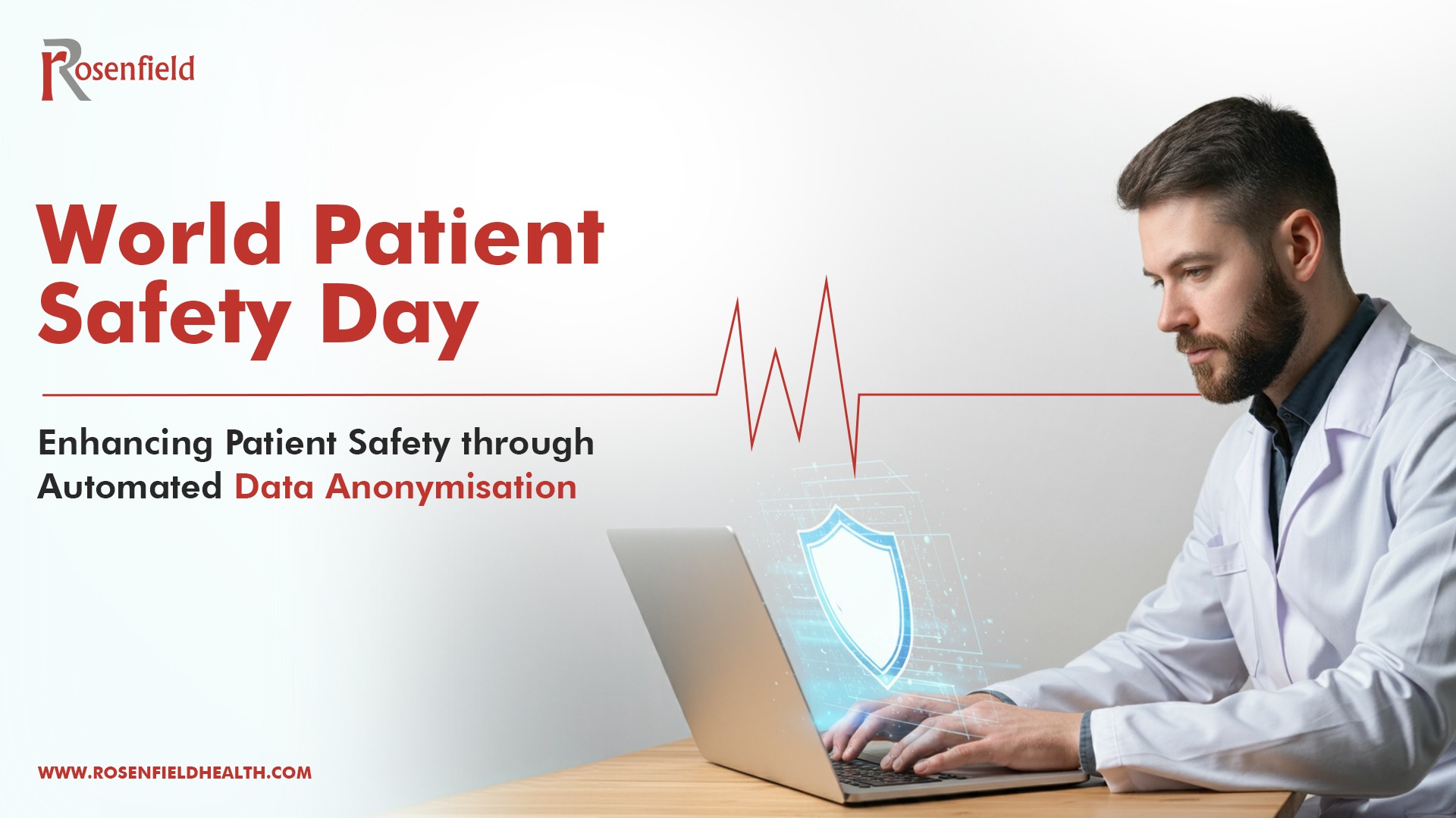
World Patient Safety Day 2024: Enhancing Patient Safety through Automated Data Anonymisation ​

Blog

World Patient Safety Day 2024: Enhancing Patient Safety through Automated Data Anonymisation ​

World Patient Safety Day 2024: Enhancing Patient Safety through Automated Data Anonymisation

World Patient Safety Day, celebrated annually on September 17th, serves as a critical reminder of the importance of protecting patients across all aspects of healthcare. This year, as we reflect on ways to enhance safety, data privacy and accuracy stand out as crucial pillars of patient care. One of the most powerful tools for ensuring these elements is data anonymisation in healthcare. 

With the growing amount of patient data generated daily, automating medical data anonymisation is no longer just an option—it’s a necessity. By leveraging automation, PACS (Picture Archiving and Communication Systems) managers can ensure patient data remains secure while significantly reducing the risk of human error. PRIX from Rosenfield Health is paving the way for automated anonymisation for patient safety, bringing new levels of accuracy, security, and efficiency to healthcare data management. 

The Need for Data Anonymisation in Healthcare 

Patient data is the lifeblood of modern healthcare. From diagnosis to treatment and research, medical data enables healthcare professionals to deliver personalised care. However, with this wealth of data comes an increased responsibility to protect patient privacy. Failure to properly anonymise patient data can lead to severe consequences, including breaches of confidentiality, identity theft, and regulatory penalties. For healthcare providers, ensuring patient confidentiality is not only a legal requirement but a moral obligation to protect those they serve. 

Data anonymisation technologies in healthcare ensure that sensitive patient information is stripped of identifying details, making it impossible to trace data back to a specific individual. But, in a complex environment like healthcare, where vast amounts of data are being processed daily, manual anonymisation is prone to error and can be highly time-consuming. 

Automation enhances both security and operational efficiency, allowing healthcare providers to process larger volumes of data quickly and accurately. It is also particularly important for organisations to remain compliant with data protection regulations such as the General Data Protection Regulation (GDPR) in the UK and Europe or the Health Insurance Portability and Accountability Act (HIPAA) in the United States. 

Automating DICOM & Pixel Data Anonymisation with PRIX 

Data anonymisation is a non-clinical task that requires great attention, taking up significant time and effort from PACS managers who would otherwise be focusing on more critical tasks. Automating this process with an innovative tool not only improves patient safety through data security and accuracy, but it also frees up healthcare professionals to focus on patient care and other high-priority tasks.  

To meet the growing demand for secure, efficient, and error-free anonymisation, Rosenfield Health developed PRIX, a cutting-edge bulk anonymiser designed to automate the anonymisation of both DICOM (Digital Imaging and Communications in Medicine) & Pixel data. By integrating this tool into healthcare workflows, PRIX enhances patient safety and boosts operational efficiency, allowing PACS managers and radiologists to concentrate on higher-priority tasks.  

Here’s how PRIX makes a difference: 

  1. Time Saving Efficiency: PRIX dramatically reduces the time required for anonymisation tasks. What traditionally takes up to 8 hours can now be accomplished in just 15 minutes. This significant reduction in time allows PACS Managers to allocate their valuable time to more critical activities, enhancing overall productivity and efficiency.  
  2. Unmatched Accuracy: By automating the anonymisation process, PRIX eliminates the risk of human error that is common with manual methods. PRIX ensures a higher level of accuracy and reliability, safeguarding sensitive information more effectively.
  3. Enhanced Focus: PRIX empowers PACS managers by freeing them from the repetitive and time-consuming task of manual anonymisation. This allows them to concentrate on more strategic and core responsibilities, significantly improving the workflow of radiologists.  

Conclusion 

As we celebrate World Patient Safety Day 2024, it’s clear that patient safety is about more than just clinical care. Protecting patient data is an integral part of safeguarding their well-being. By leveraging the power of automating medical data anonymisation, healthcare providers can enhance both privacy and accuracy, ensuring that patient data remains secure without compromising on efficiency. 

With PRIX, Rosenfield Health is at the forefront of providing automated anonymisation for patient safety, offering a solution that is not only efficient but also reliable and compliant with global standards. This World Patient Safety Day let’s commit to improving patient safety through innovative solutions like PRIX that make healthcare data management safer and more accurate for everyone—while giving radiologists and PACS managers more time to focus on what truly matters: patient care. 

#RosenfieldHealth #WorldPatientSafetyDay2024 #DataAnonymisation #HealthcareDataSecurity #AutomatedAnonymisation #PRIX #PatientSafety #DataAccuracy  


We are certified: ISO/IEC 27001:2022 Award

Blog

We are certified: ISO/IEC 27001:2022 Award


Rosenfield Health has officially achieved ISO/IEC 27001:2022 certification!

Thanks to the great efforts and dedication of Rosenfiled Health’s team, we are ISO 27001/2022 certified!

This significant milestone reflects our responsibility towards our success partners to safeguard confidential data and maintain patients’ privacy. Our team has worked tirelessly to meet these international standards and embraced the challenge to demonstrate exceptional reliability and commitment.

Our partners can trust that any sensitive data is in safe hands.

Case Study: The EMRAD Journey with iCode REALM ​

Blog

Case Study: The EMRAD Journey with iCode REALM ​

Case Study: The EMRAD Journey with iCode REALM

Background 

The East Midlands Radiology Consortium (EMRAD) is a network aiming to deliver expert radiology services across the region. However, the REALM chairs within EMRAD faced overwhelming workloads, making the management of REALM meetings a significant challenge. From anonymising studies to recording learning points and summarising meetings, the process can be time-consuming. 

The Challenge 

REALM meetings are crucial for continuous learning in radiology, allowing professionals to learn from discrepancies and successes. The manual effort required to manage these meetings was substantial, consuming valuable time and resources that could otherwise be directed towards patient care. 

The iCode REALM Solution 

iCode REALM was introduced as a vendor-neutral solution seamlessly integrated into the PACS (Picture Archiving and Communication Systems) environment, designed to facilitate the execution of REALM meetings in accordance with RCR guidelines. 

With iCode REALM, EMRAD experienced a transformation in their workflow: 

  1. Streamlined Automated Workflow: The process of managing and running REALM meetings became more straightforward and efficient. 
  2. Anonymisation of Cases: Patient and physician data were anonymised, maintaining privacy, and focusing on learning from case content, and facilitating discussions around discrepancies in a blame-free environment. 
  3. Time and Resource Efficiency: The automated system reduced the manual effort required, freeing up time for REALM chairs. 
  4. Reduced Workload Challenges: REALM chairs could focus more on their primary responsibilities rather than administrative tasks.
  5. Powerful Meeting Management Tool: iCode REALM provided a robust platform for scheduling, executing, and reviewing meetings.
  6. Analytics and Reporting: The system offered insights into meeting outcomes, helping to enhance quality and processes. 

In conclusion, the implementation of iCode REALM at EMRAD led to a more efficient and effective learning environment, revolutionising radiology practice and raising the bar for patient care and safety. 

To learn more about how iCode REALM can transform your radiology practice, visit iCode REALM. 

From Chaos to Clarity: Managing Critical Result Escalations with iCode CRN

Blog

From Chaos to Clarity: Managing Critical Result Escalations with iCode CRN

From Chaos to Clarity: Managing Critical Result Escalations with iCode CRN

Introduction: In the high-stakes world of radiology, where seconds matter, the management of critical results can be chaotic. Radiologists and referring physicians find themselves caught in urgent scenarios, striving to ensure patient safety while navigating complex workflows. This is where iCode Critical Results Notification (CRN) steps in—a powerful solution that transforms chaos into streamlined communication and accountability. 

The Chaotic Landscape 

Imagine this following scenario: A radiologist recognizes a critical finding—a life-threatening condition like a rupture, a stroke, or an urgent abnormality. The time starts to run. The pressure to accurately and swiftly convey this information to the referrers is overwhelming. In the absence of a streamlined system, this process can be prone to errors, delays, and miscommunication, risking patient safety and outcomes. Radiologists suffer from: 

  1. Manual Processes: Radiologists manually make phone calls or send emails to referring physicians.
  2. Fragmented Systems: Patient data exists in disparate systems—Picture Archiving and Communication Systems (PACS), Radiology Information Systems (RIS), and Electronic Health Records (EHR). Coordinating across these silos can be stressful. 
  3. Accountability Challenges: Who acknowledged the critical result? Was it delivered promptly? Lack of documentation and record keeping can lead to accountability gaps.

Introducing iCode CRN: A Beacon of Clarity 

iCode CRN appears as a beacon of clarity in this chaotic landscape. Let’s explore how it revolutionizes critical result management: 

  1. Unified Communication Hub: No more toggling between screens; everything is at the radiologist’s fingertips. iCode CRN integrates seamlessly with existing systems (PACS, RIS, EHR) to obtain patient details, study information, and referring physician contacts.
  2. Urgent Result Communication: Through push notifications, SMS, HL7 messages, and email, iCode CRN ensures that critical results reach the right referrer promptly. No more lost emails or missed calls.  
  3. Radiologist Accountability: iCode CRN tracks every step. Radiologists receive delivery confirmations, and referrers acknowledge receipt. Accountability becomes transparent.
  4. Patient Safety First: By automating processes, iCode CRN frees radiologists to focus on clinical interpretation, preventing crucial information from slipping through the cracks.
  5. Mobile Empowerment: The iCode CRN mobile app allows radiologists to access and acknowledge critical results on the go. 

Conclusion: From chaos to clarity—the journey is now guided by iCode CRN. Radiologists wield a powerful tool that not only streamlines workflows but also safeguards lives. As the critical result escalates, so does our commitment to patient safety. 

3 Tips to Organise an Effective Tumour Board

3 Tips to Organise An Effective Tumour Board

Blog

3 Tips to Organise An Effective Tumour Board

Enhancing Cancer Care: 3 Tips to Organise an Effective Tumour Board

Introduction: As we mark World Cancer Month, it’s a timely reminder of the global impact of Cancer and the importance of effective collaboration in combating this disease. It’s important to reflect on the critical role that well-organised Tumour Board meetings play in enhancing cancer care efficiency. These multidisciplinary gatherings bring together healthcare professionals from various specialties to collectively review complex cases, discuss treatment options, and formulate comprehensive care plans for cancer patients. By fostering collaboration, innovation, and evidence-based decision-making, these Board meetings contribute significantly to improving patient outcomes and quality of life. In this article, we’ll explore three tips to organise Tumour Boards effectively and enhance cancer care outcomes. 

1. Establish Clear Objectives and Agendas

Set clear objectives and agendas for each meeting to ensure focus and productivity. Define specific goals, such as reviewing diagnostic imaging, discussing treatment options, and developing individualised care plans.

2. Enhance Data Accessibility and Integration

Access to comprehensive patient data is crucial in discussions and decision-making. Integrate electronic health records (EHRs), imaging studies, pathology reports, and other relevant information into a unified platform. By centralizing data access, healthcare professionals can make more informed decisions, reduce duplication of efforts, and improve patient outcomes.

3. Ensure All Images Are Reviewed Prior to the Tumour Board

Comprehensive imaging studies are a cornerstone of cancer diagnosis and treatment planning. It is also one of the standards the RCR addressed in their recent article. To maximize the effectiveness of Tumour Board meetings, it’s essential to ensure that all relevant images are thoroughly reviewed beforehand. This includes radiographs, CT scans, MRIs, PET scans, and any other imaging modalities pertinent to the patient’s case. By meticulously examining these images prior to the meeting, healthcare professionals can identify key findings, gain a deeper understanding of the patient’s condition and save time during the meeting.

In today’s digital age, leveraging technology can significantly enhance the efficiency and effectiveness of Tumour Board meetings. With solutions like iCode MDT, healthcare professionals can streamline the entire process, from adding comments prior to the meeting to documenting patient data and treatment plans. By using iCode MDT’s intuitive platform, organizers can create customized agendas, assign tasks, and track progress seamlessly. Additionally, iCode MDT facilitates real-time collaboration and communication among team members, ensuring that everyone is on the same page and working towards common goals. Incorporating technology like iCode MDT into Tumour Board meetings not only saves time and resources but also improves overall outcomes for cancer patients.  

Conclusion: Organising Tumour Boards effectively is essential in enhancing cancer care and improving patient outcomes. By fostering reviewing images, establishing clear objectives and enhancing data accessibility, healthcare professionals can maximize the impact of MDT meetings. Leveraging innovative solutions like iCode MDT further enhances collaboration, streamlines processes, and ultimately contributes to better cancer care delivery. Together, let’s continue to work towards a future where cancer is no longer a life-threatening disease, but a manageable condition with improved treatment options and outcomes.