Radiology Teaching Files

Radiology Teaching Files - Rosenfiled Health

Radiology Teaching Files

Radiology teaching files are powerful tools for radiologists. They allow the archiving of interesting cases to ensure important findings are not missed while also enhancing diagnostic skills.

These teaching files serve multiple purposes, whether it’s for educating radiology students, discussing cases with colleagues, contributing to the medical community, or keeping up with effective e-learning methods for radiology.

What Are Teaching Files?

Radiology teaching files are collections of clinical cases with teaching value.

These files provide real examples of radiology cases that may help with teaching purposes, whether for radiologists or radiology students.

Generally, radiology education relies on studying and referencing previous patient cases but storage, accessibility, and shareability with students can present challenges. Additionally, studies should be anonymized.

So, radiology teaching files can serve several purposes, such as:

  • To ensure important findings are not missed.
  • Archiving cases for clinical follow-up.
  • As a reference for better understanding diseases.
  • Facilitating secure Knowledge transfer and assessment.
  • Enabling quick retrieval of anonymized patient cases.

Structure Of Radiology Teaching Files

Radiology teaching files are organized to make them easy for learners to navigate and understand. Each case typically includes:

  • Patient information: This Includes the patient’s age, gender, medical history, and symptoms.
  • Imaging studies: X-rays, CT scans, MRIs, or ultrasounds are often presented in multiple views to better understand the cases.
  • Annotations and labels: Key areas of the image are often highlighted to direct the learners to a significant finding.
  • Diagnosis and discussion: A detailed explanation of the diagnosis, including why certain findings on the images led to that conclusion.
  • Teaching points: Summary points that highlight important lessons from the case.

Teaching Files Types

Teaching files can be divided into three types:

1. Personal Teaching Files

Personal teaching files are for individual use. The owner may have an interest in a very specific case for quality control, reviewing his work, teaching, or clinical follow-up.

2. Shared In-House Teaching Files

Personal teaching files can be migrated into a shared teaching file environment. Additional information can be added to make the case available for colleagues to view and learn from.

3. Public Teaching Files

They are built on a shared teaching files model but contain more comprehensive content. They may undergo a formal review before publication and sometimes require a subscription fee.

Teaching File Case Types

Teaching file case types include:

1. Cardiac

Cardiac teaching files focus on heart-related cases and cardiovascular conditions. They include imaging for:

  • coronary artery disease
  • congenital heart anomalies
  • heart failure
  • valvular diseases
  • others

Radiologists can use these files for better understanding of heart structure and function. Files can be also helpful for assessment of cardiac conditions.
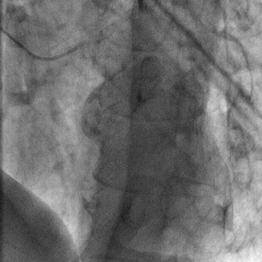
Cardiac and Interventional teaching files

2. Chest

Chest teaching files cover diseases of the chest, such as:

  • pneumonia.
  • chest tumors.
  • interstitial lung diseases.
  • pleural abnormalities.

They help radiologists enhance their skills in chest cases.

3. Gastrointestinal

Gastrointestinal teaching files focus on the digestive system including the esophagus, stomach, intestines, liver, pancreas, and biliary system.

They include cases of various gastrointestinal diseases, such as:

  • Liver diseases
  • Colorectal cancers
  • pancreatitis
  • bowel obstruction

Radiologists can archive these teaching files for educational purposes or to enhance their skills.

4. Genitourinary

These files focus on the urinary and reproductive systems. Files can include:

  • Renal cell carcinoma
  • Polycystic kidney disease
  • Urinary tract infections
  • Urinary stones
  • Hydronephrosis
  • Ovarian masses

These files help radiologists assess urological structure and functions and improve their experience in the diagnosis of urinary tract masses and pathologies.
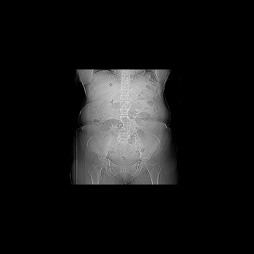
CT- Genitourinary teaching files

5. Interventional

Interventional teaching files include some procedures, such as:

  • Biopsies
  • Catheter placements
  • Angioplasty
  • Tumor ablation

These cases help radiologists learn more about the procedures and improve patient management skills.

6. Mammography

Mammography files specialize in breast imaging cases, such as:

  • Breast tumors
  • Fibrocystic changes
  • Breast cysts
  • Ductal carcinoma in situ

Radiologists can gain valuable experience in breast cases through mammography teaching files.
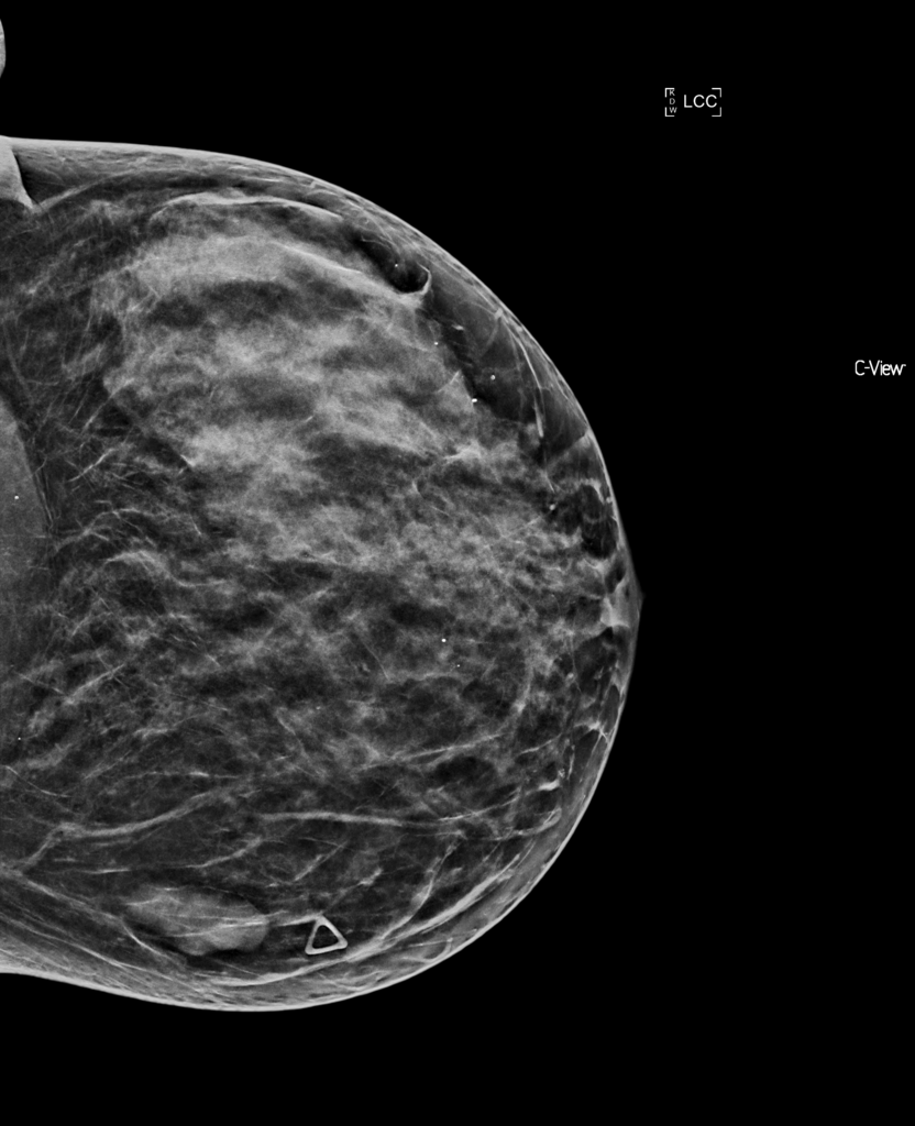
Mammography teaching files

7. Musculoskeletal

Musculoskeletal teaching files focus on improving radiologist’s skills in recognizing bone and soft tissue abnormalities, such as:

  • Osteoarthritis
  • Soft tissue tumors
  • Fractures
  • Bone tumors
  • Sports-related injuries

8. Neuroradiology

Neuroradiology files guide radiologists in recognizing brain abnormalities and how to differentiate normal from abnormal brain or nervous structure. They cover cases, such as:

  • Stroke
  • Brain tumors
  • Multiple Sclerosis
  • Traumatic brain injuries

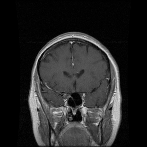
MRI Neuroradiology teaching files

9. Nuclear Medicine

These files cover radiotracer use, interpretation of uptake patterns, and correlation with clinical scenarios. They provide cases like:

  • Thyroid nodules
  • Bone metastases
  • Neuroendocrine tumors

10. Pediatrics

Pediatric files train radiologists to recognize specific conditions in children. Pediatric cases often include:

  • Rickets
  • Congenital malformations
  • Neuroblastoma
  • Pediatric trauma
  • Respiratory conditions

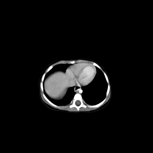
Pediatric teaching files

11. Ultrasound

Ultrasound case studies are essential for helping radiologists identify numerous conditions and enhance their diagnostic skills. They may include cases on:

  • Gallstones
  • Liver disease
  • Obstetric and gynecologic conditions
  • Thyroid nodules
  • Abdominal conditions
  • Vascular conditions

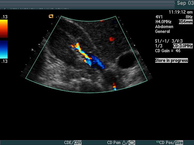
Ultrasound teaching files

Importance Of Teaching Files For Radiologists

Radiologists may be advised to build their teaching files for the following reasons:

1. Increase Your Expertise

Establishing teaching files encourages radiologists to engage with cases on a deeper level.

It fosters critical thinking in analyzing images, making accurate diagnoses, and explaining findings.

This process sharpens diagnostic skills and enhances a radiologist’s level of experience.

2. Education Purposes

Radiology teaching files enrich the medical community by providing real and often interesting cases. This helps medical students, radiologists, and colleagues. Importantly, difficult cases recorded in teaching files will be more easily diagnosed in the future.

3. Self-Learning And Review

Teaching files are not only for the broader medical community but can also serve as a valuable personal learning resource. Having cases documented allows radiologists to review challenging cases, rare pathologies, or those with subtle findings.

iCode Teaching Files

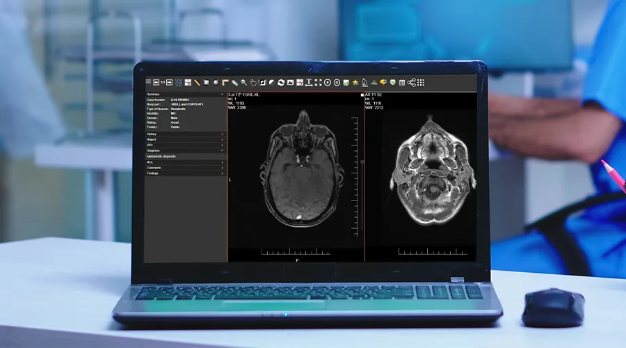
iCode Teaching Files is a web teaching files solution that helps radiologists archive their interesting studies and build imaging libraries according to ACR standards.

It archives interesting cases on a separate server with a separate database. You can keep the radiology teaching archive safe when the PACS vendor is changed.

Examples of iCode teaching files features include:

  • Easy file search and retrieval
  • Robust data anonymisation
  • Ability to export studies with several media files, such as presentations, videos, JPEG, TIFF & DICOM..etc
  • Manage teaching events easily with a streamlined workflow and lectures bank to enhance learning
  • Building scalable teaching archive across specific region or country.

iCode Teaching Files Radiology Exam System

The iCode Teaching File Solution is revolutionizing radiology education with its comprehensive exam module, designed to enhance both teaching and learning.

This system assists radiologists in creating radiology exams for their students. The exam module in iCode Teaching Files allows radiologists to craft multiple-choice questions (MCQs) based on archived medical images.

It also enables educators to share quiz links with their students, allowing them to take exams directly through the teaching files system.

With detailed analytics and instant feedback, both learners and educators can track progress and identify areas for improvement, ultimately elevating the quality of training.

iCode offers a rich set of features tailored to meet your educational needs, enabling you to archive cases quickly and effortlessly. Additionally, its comprehensive exam module supports and enhances the educational process.

DICOM Anonymisation Software

DICOM Anonymisation Software

DICOM Anonymisation Software

DICOM anonymisation software is essential for the secure storage and sharing of medical images. It preserves patient privacy, ensures that personal data is anonymised and facilitates the use of images for teaching or research purposes without exposing sensitive patient information.

How do you choose the best DICOM anonymisation software? This article will discuss the key considerations for this.

What is DICOM anonymisation software?

DICOM anonymisation software protects medical images privacy by removing or replacing personal information within DICOM files.

DICOM anonymisation software’s goal is to make the data anonymous, allowing images to be used for medical or research purposes without risking patient privacy.

For example, when doctors share radiological images with colleagues, they must remove all identifiable information, such as the patient’s name, address, or other personal details.

How does it work?

First, DICOM files are uploaded into the anonymisation software, which then scans the files for metadata tags containing personal information.

The software then removes or replaces this information with generic placeholders to protect the patient’s identity.

Pros of DICOM anonymisation software

DICOM anonymisation software is important to protect patients information especially with growing use of electronic health records that lead to more data being shared, increasing chances for accidental leaks of information.

Some benefits of DICOM anonymisation software include:

1. Compliance with regulations

Anonymising patient data is a legal requirement to protect patient confidentiality, so following strict privacy regulations, such as GDPR or HIPAA is essential.

2. Keeping patients’ information secure

Protecting medical information of the patients is more critical than ever, as cyberattacks have become sophisticated and can cause serious harm by stealing patients information or making it vulnerable to malicious actors.

3. Increase collaboration

Anonymised DICOM files help with collaboration across different departments, healthcare facilities, and research centers without compromising patient’s privacy.

4. Batch processing

Many DICOM anonymisation tools support batch processing, allowing the anonymisation of large sets of images at once (bulk DICOM anonymisation).

This feature is especially helpful for large healthcare facilities and research centers that handle substantial volumes of medical images.

Cons

Although bulk DICOM anonymisation software are very useful, the may have some cons:

1. Complex to use

Some DICOM anonymisation software can be complex to use, especially for healthcare professionals who are less technically proficient.

As a result, it may require some training, or having technical support to use DICOM anonymisation software properly without affecting the quality of patient care.

2. Potential for human error

Although anonymisation software can automate the process, there’s always a risk of human error.

For instance, a user may accidentally select the wrong settings, causing improper anonymisation, which results in unintentional patient data exposure.

3. Loss of data integrity

Sometimes anonymisation software can cause a loss of certain non-essential data that could be important in specific situations.

A clear example of this is when removing all patient-related data, it’s hard to trace an image back to the original patient for follow-up.

4. Not always 100% accurate

DICOM Anonymiser tools are very effective, although some advanced DICOM metadata might not be anonymised properly, which leaves some sensitive information exposed.

As a result, it may be essential to check the anonymisation process periodically to ensure patient privacy.

How to choose the right DICOM anonymisation software?

Choosing the best DICOM anonymisation software for your practice depends on many factors:

1. Ease of use

It’s preferable to look for a user-friendly software that can integrate easily into your workflow. As a healthcare provider, you should seek software with minimal learning time, ensuring ease of use without disrupting workflow.

2. Complaint with regulations

you have to ensure that the software you choose complies with privacy laws to avoid any legal problems and protect patient privacy.

3. Security features

DICOM anonymisation software varies in the security features, so you should make sure to choose the software that provides high protection of data during storage and transmission.

Some security features, such as encryption and secure cloud storage, provide great protection for patient privacy.

4. Scalability

The volume of the data may differ from healthcare facility to another, so you have to ensure that you have a software that can handle a large volume of data, whether now or later in case you need that.

This is very important especially for healthcare facilities or research centers that deal with a large amount of medical images and they sure need to protect their patient privacy.

5. Little errors

Although 100% accuracy is impossible, look for DICOM anonymisation software with minimal errors to ensure maximum security of patient information and prevent unintended data leaks.

6. Integration with other systems

A perfect DICOM anonymisation software should integrate well with other systems, such as the PACS system.

Best DICOM anonymisation software

PRIX Bulk DICOM Anonymiser is a powerful tool to bulk anonymise medical images and can integrate with PACS system.

It has some features that fit your need:

  • Effortless anonymisation: PIXR Bulk DICOM Anonymiser can anonymise large volumes of studies at once.
  • Multiple anonymisation profiles: It customizes with multiple profiles to meet the specific needs of different research, teaching, and clinical trials.
  • DICOM and pixel data anonymisation: It ensures precise anonymisation of the patient data in US images, maintaining data integrity and patient privacy with zero errors.
  • Integration with other systems: PRIX integrates with PACS, clinical trial servers, providing detailed log for each anonymisation step.

You can find what you need for anonymising your studies and cases perfectly in PIXR Bulk DICOM Anonymisation with its unique features.

FAQs

What is DICOM software?

DICOM software refers to specialized tools and applications designed to view, store, and share medical images in DICOM format, the standard format used in medical imaging.

How to anonymise DICOM files?

You can anonymise DICOM files using DICOM anonymisation software, as it preserves patient privacy while using images in teaching, research, or other uses.

PACS System Radiology

PACS System Radiology

PACS system radiology

PACS system radiology is a secure, reliable method to store, retrieve, and transport medical images. It provides benefits, such as remote access, and better-organized patient data.

They eliminate the need for hard-copy films and use digital images instead. This can help with providing better patient care and making more accurate diagnoses, especially in radiology.

What is PACS?

PACS stands for Picture Archiving and Communication System. It securely transports medical imaging information, allowing healthcare professionals to store and transmit images and clinical reports.

How does it work?

The PACS system stores and transfers medical images in DICOM (Digital Imaging and Communications in Medicine) format and supports several functions:

  • Image acquisition: Medical images are digitally captured from multiple imaging methods, such as X-ray, CT, and MRI, adhering to DICOM standards.
  • Image transfer and storage: These digital images are sent to the PACS server that stores and archives them in a central database.
  • Image distribution and viewing: Radiologists can access and view stored images from any PACS workstation connected to the network.
  • Reporting: Radiologists can create and save diagnostic reports with their images in the PACS.
  • Integration: PACS systems can integrate with other systems, such as RIS (Radiology Information System).

PACS and RIS

PACS can integrate with the Radiology Information System (RIS), enabling bi-directional communication between the systems. RIS sends patient data to PACS, and PACS returns acquired images and reports, allowing a coordinated radiology workflow.

Importance of PACS in radiology

Radiologists benefit from PACS in many ways, including:

  • Remote access: PACS enables radiologists to access diagnostic images anytime, improving diagnosis accuracy, speed, and healthcare quality.
  • Film archives replacement: PACS eliminates the need for film archives, reduces physical storage requirements, and promotes efficient cooperation across departments.
  • Easy for radiologists: Radiologists can use the PACS system to manage their work. They take the necessary images, review them in the workstation, sending them to the digital archive, and the images are now available for healthcare professionals who have access to the PACS system.

Types of PACS systems

Types of PACS systems include:

1. Traditional PACS

Traditional PACS represents the cornerstone of image storage, retrieval, and distribution within a local network.

They offer strong control and privacy but it’s limited by physical space and requires continuous maintenance.

Traditional PACS are perfect for facilities that prefer keeping all operations in-house and have the necessary infrastructure.

2. Cloud-based PACS

They are more modern than traditional PACS, as they benefit from the internet for image storage and management. The images can be accessed from any device, at any time without great concern about physical storage limitations or maintenance.

They offer scalable storage solutions, usually requiring a subscription.

3. Hybrid systems

Hybrid systems blend traditional and cloud-based PACS. They offer the best of both systems:

  • Control and Security
  • flexibility and scalability of the cloud

They are fit for those looking forward to maintaining on-site backup while taking advantage of cloud storage as well.

Benefits of PACS system radiology

Medical PACS systems can provide a lot of benefits, especially in the radiology field. Some of these benefits include:

  • More efficient diagnoses
  • Reduced costs
  • High-quality medical images
  • Instant remote access for medical images
  • Increased integration between departments and facilities
  • Improve patient care
  • Ease of use and scalability of cloud-based PACS

For more details about some of the benefits:

  • Reduced costs: Digital storage is generally less expensive than storage needed for hard-copy films, which makes the PACS system a cost-effective solution.
  • Better patient data management: All patient information can be accessed through a single point of access as images are integrated with the radiology information system.
  • Faster workflow: There’s no need for manual transport and retrieval of images. It’s a rapid process with the help of the PACS system, as radiologists can access images quickly and provide better patient care.

Best PACS system radiology

The best PACS system according to the number of installs includes:

  1. GE Healthcare – Centricity
  2. Change Healthcare – Radiology Solutions
  3. FUJIFILM Medical Systems – Synapse
  4. IBM – Merge PACS
  5. Philips Healthcare – iSite
  6. Agfa Healthcare – Impax
  7. Novarad – NovaPACS
  8. Sectra – SectraPACS
  9. Carestream Health – Carestream vue
  10. Change Healthcare

PACS system radiology cost factors

How much does a PACS system cost? There are many factors affecting the cost of the PACS system, such as:

  • Total storage capacity: Solid-state storage (SSD) is more available and much faster to access than spinning disks (HDD). SSD costs about double the similar capacity of disk storage.
  • Maintenance and support: The PACS system requires annual maintenance and support which may affect the cost of the PACS system.
  • Type of PACS system used: Also choosing the appropriate type of PACS system for you may influence the total cost.

FAQs

What is the PACS system in radiology?

PACS system is a secure method to store and transport medical images according to DICOM format. It helps radiologists to access diagnostic images anywhere, sharing images with other healthcare professionals, and providing better patient care.

What are the main components of PACS?

PACS system for radiology components include:

  • Main server: The main server is the most important part of a PACS. It includes the database structure, RIS interface, web servers, and other image distribution servers and interfaces.
  • Database: This refers to the place at which all the important information is stored including all the data related to the patient study and examination.
  • Health level seven (HL7): The HL7 is the part that receives all the info caused by RIS and then forwards it to the PACS.
  • Imaging modalities: These are machines used to capture digital medical images, such as X-ray machines, MRI machines, and Ultrasound equipment.
  • Workstations: These are computers equipped with DICOM viewers that allow radiologists to access, view, and interpret stored images.
  • Storing archives: DICOM images are stored in the servers or cloud storage, either on-site, in the cloud, or both (hybrid).

A secure network connection is also necessary for the healthcare PACS system to work efficiently.

Rosenfield Exhibiting at UKIO 2024

News

Rosenfield Exhibiting at UKIO 2024

Rosenfield at UKIO 2024

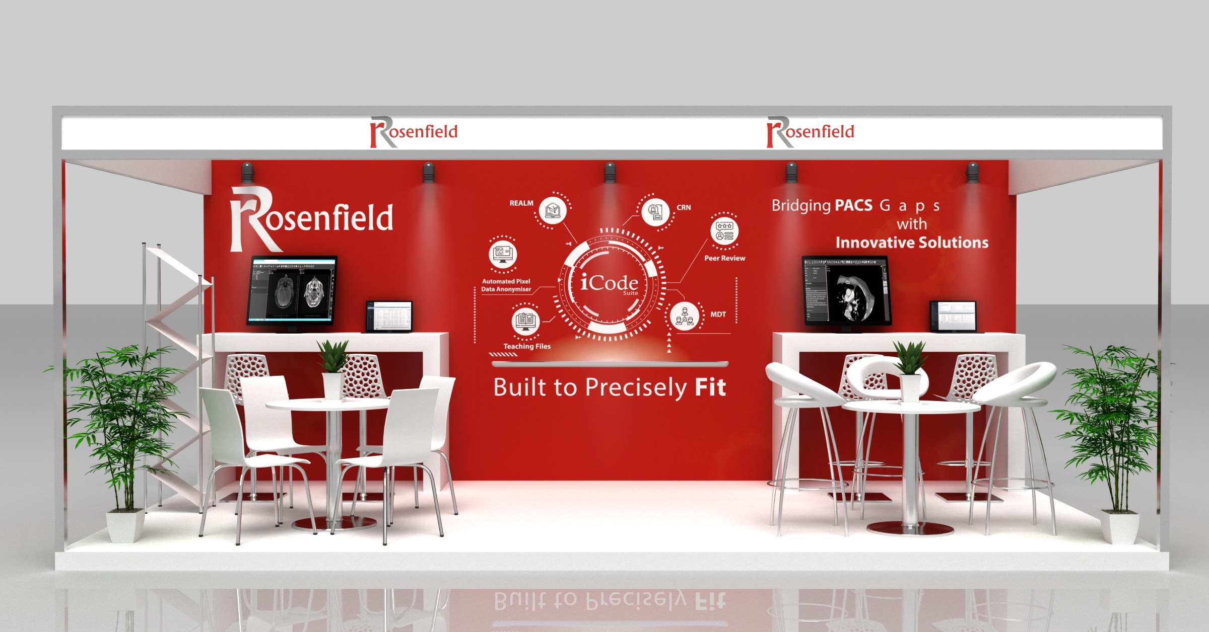
We’re thrilled to announce that we will be exhibiting at hashtagUKIO24!

Join us to get hands-on with our latest innovative solutions and dive into our interactive demos! Our team is ready to meet you and discuss how our products can empower your workflow. 

Mark Your Calendars:
Date: 10,11,12 June, 2024
Location: ACC, Liverpool
Booth: A5/10


Fill out this form to schedule your Demo during the event:
https://lnkd.in/drKPmvwu

We can’t wait to see you there and explore the possibilities together

#UKIO #Medical_imaging #HealthcareIT #Oncology #Radiology